Anatomy and Physiology Lab Manuals are crucial resources, often openly licensed, designed to complement coursework for nursing and allied health students.
These manuals, like those by Allen, Harper, Marz, and others, provide practical exercises and guidance for understanding complex biological concepts.
They are available in various formats, including PDF and ePUB, facilitating accessible learning and skill development in anatomical studies.
Purpose of Lab Manuals
Anatomy and Physiology Lab Manuals serve a vital purpose in reinforcing theoretical knowledge through hands-on application. They bridge the gap between classroom learning and practical skills essential for future healthcare professionals.
These manuals, such as those utilized in nursing and allied health programs, provide structured exercises – dissections, microscopy, and data analysis – to deepen understanding of bodily structures and functions.
They guide students through the scientific method, fostering experimental design and data interpretation abilities. Furthermore, manuals like those authored by Connie Allen and others, offer step-by-step procedures, ensuring accurate technique and safe laboratory practices.
Ultimately, the core purpose is to cultivate competency and confidence in applying anatomical and physiological principles, preparing students for clinical settings.
Importance in Nursing & Allied Health
Anatomy and Physiology Lab Manuals are fundamentally important for students pursuing careers in nursing and allied health professions. A strong grasp of anatomical structures and physiological processes is critical for accurate patient assessment, diagnosis, and treatment.
These manuals, like the one described as “great” from the Open Textbook Library, provide the practical experience needed to translate theoretical knowledge into clinical competence; Skills honed through dissections and analyses directly impact patient care.
Understanding how organ systems interact, as explored in these labs, is essential for recognizing abnormalities and implementing effective interventions. Access to resources, even in digital formats like PDF, ensures widespread learning opportunities.
Ultimately, proficiency gained through lab manuals contributes to safer, more effective healthcare delivery.

Essential Lab Equipment & Techniques
Anatomy and Physiology Lab Manuals guide students through utilizing essential tools like microscopes and dissection instruments for effective study and analysis.
Microscopy
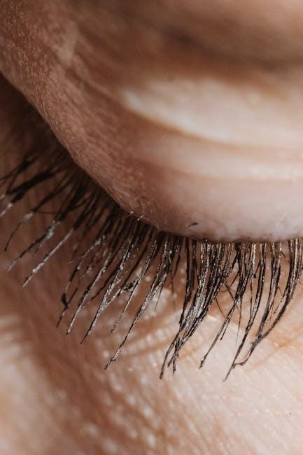
Microscopy is a foundational technique in Anatomy & Physiology labs, heavily emphasized within lab manuals. Students learn to prepare slides, focusing on tissue samples for detailed observation.
Lab manuals provide step-by-step instructions for proper microscope usage, including focusing techniques and identifying various magnification levels.
Understanding microscopic structures – cells, tissues, and their components – is vital for comprehending organ system functions.
These manuals often include diagrams and exercises to aid in identifying key features of cells and tissues under the microscope.
Proper slide preparation and microscope handling are crucial skills developed through these practical exercises, ensuring accurate observations and analysis.
This skill is essential for future healthcare professionals.
Dissection Tools & Procedures
Dissection, a core component of Anatomy & Physiology labs, is thoroughly covered in lab manuals, detailing proper tool usage and safety protocols.
Manuals illustrate and explain the function of instruments like scalpels, forceps, and scissors, emphasizing careful handling to preserve specimen integrity.
Step-by-step dissection procedures, often utilizing fetal pigs or other specimens, are provided with detailed diagrams for accurate identification of structures.
Students learn to systematically expose and examine organs, understanding their anatomical relationships within the body.
Lab manuals stress the importance of precise incisions and careful tissue separation for optimal visualization and learning.
These practical skills build a strong foundation for anatomical knowledge.
Safety Protocols in the Lab
Anatomy & Physiology Lab Manuals prioritize safety, dedicating sections to essential protocols for a secure learning environment.
These manuals outline proper handling of specimens, including disposal procedures to prevent contamination and ensure biohazard safety.
Detailed guidelines cover the use of dissection tools, emphasizing careful techniques to avoid self-injury and damage to specimens.
Personal Protective Equipment (PPE), such as gloves and goggles, is consistently highlighted as mandatory for all lab activities.
Emergency procedures, including first aid and spill cleanup, are clearly explained to prepare students for unforeseen events.
Adherence to these protocols is crucial for responsible and effective laboratory practice.

Body System Labs: Overview
Anatomy & Physiology Lab Manuals systematically explore body systems – skeletal, muscular, nervous, cardiovascular, and more – through dissections and analyses.
These labs enhance understanding of organ structure and function, vital for allied health professions.
Skeletal System Lab
Skeletal System Labs, as detailed in Anatomy and Physiology Lab Manuals, are foundational for understanding the body’s structural support and movement.
These labs emphasize bone identification, requiring students to accurately name and locate various bones within the human skeleton, utilizing provided diagrams and specimens.
A key component involves analyzing bone structure, examining features like compact and spongy bone, epiphyses, and diaphyses to comprehend their functional roles.
Students learn to differentiate bone types – long, short, flat, irregular – and correlate their shapes with specific functions.
Practical exercises often include identifying bone markings, such as foramina, processes, and tubercles, which serve as attachment points for muscles and ligaments.
These labs are essential for building a strong base in anatomical terminology and skeletal anatomy.
Bone Identification & Structure
Bone Identification & Structure exercises within Anatomy and Physiology Lab Manuals are critical for mastering skeletal anatomy.
Students utilize lab resources to accurately identify individual bones – like the femur, humerus, and skull – based on their unique morphological features.
Labs focus on distinguishing between axial and appendicular skeletons, understanding the organization and function of each division.
Detailed examination of bone structure reveals the composition of compact and spongy bone, and the role of osteons in providing strength.
Students learn to identify key anatomical landmarks, including processes, foramina, and fossae, and their significance for muscle attachment and nerve passage.
These exercises build a foundational understanding for further study of the musculoskeletal system.
Muscular System Lab
Muscular System Labs, as detailed in Anatomy and Physiology Lab Manuals, provide hands-on experience with muscle tissues and their functions.
Students learn to differentiate between the three muscle tissue types – skeletal, smooth, and cardiac – based on microscopic examination and physiological properties.
Labs emphasize identifying major skeletal muscles, their origins, insertions, and actions, often utilizing models or diagrams.
Understanding muscle fiber arrangement and its impact on force production is a key learning objective.
Practical exercises may involve palpating muscles on anatomical models or even on themselves, to understand their location and function.
These labs solidify comprehension of muscle contraction mechanisms and their role in movement.
Muscle Tissue Types & Function
Anatomy and Physiology Lab Manuals dedicate significant sections to exploring Muscle Tissue Types & Function, crucial for understanding bodily movement.
Labs focus on distinguishing skeletal muscle – responsible for voluntary movements – from smooth muscle, found in internal organs, and cardiac muscle, exclusive to the heart.
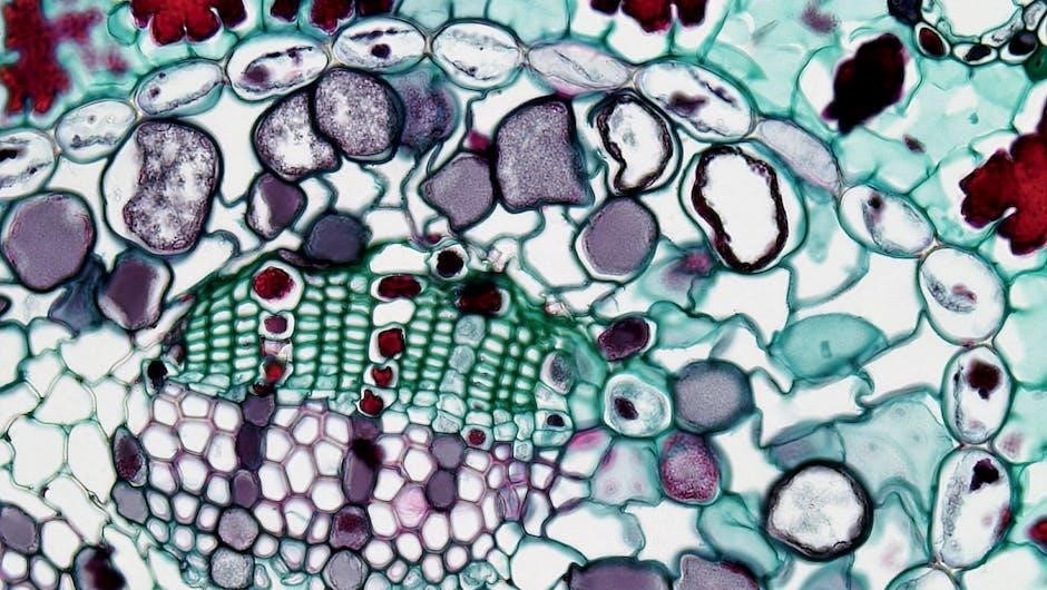
Microscopic slides allow students to observe the unique structural characteristics of each type, like striations in skeletal and cardiac muscle.
Exercises explore how muscle contractions generate force, relating structure to function.
Understanding the role of sarcomeres, actin, and myosin is emphasized.
Labs often include activities demonstrating the physiological differences in contraction speed and endurance between muscle types.
Nervous System Lab
Anatomy and Physiology Lab Manuals dedicate a substantial portion to the Nervous System Lab, a cornerstone of understanding bodily control and response.
These labs commonly feature Brain Dissection & Neuroanatomy, allowing students to physically identify key structures like the cerebrum, cerebellum, and brainstem.
Manuals guide students through tracing neural pathways and understanding the functional areas of the brain.
Activities often involve examining prepared slides of nervous tissue to observe neurons and glial cells.
Labs explore reflex arcs and synaptic transmission, demonstrating the speed and efficiency of nervous system communication.
Understanding the organization of the central and peripheral nervous systems is a key objective.
Brain Dissection & Neuroanatomy
Brain Dissection & Neuroanatomy are central components within Anatomy and Physiology Lab Manuals, offering hands-on learning experiences.
Students meticulously dissect sheep brains, a common practice due to their structural similarity to the human brain, guided by detailed manual instructions.
Labs focus on identifying major brain regions – cerebrum, cerebellum, brainstem – and their associated functions.
Manuals aid in tracing neural pathways and understanding the lobes of the cerebrum (frontal, parietal, temporal, occipital).
Neuroanatomical studies emphasize the importance of structures like the hippocampus and amygdala.
Students learn to correlate anatomical structures with physiological processes, enhancing comprehension of neurological function.
Cardiovascular System Lab
Cardiovascular System Labs, detailed within Anatomy and Physiology Lab Manuals, provide practical insights into circulatory function.
A key component is often heart dissection, allowing students to identify chambers, valves, and major blood vessels like the aorta and vena cava.
Labs incorporate blood analysis, examining blood components and performing tests like blood typing and hematocrit determination.
Manuals guide students through tracing blood flow through the heart and systemic/pulmonary circuits.
Physiological measurements, such as pulse rate and blood pressure, are routinely taken and analyzed.
Students correlate anatomical structures with physiological processes, understanding how the heart’s structure supports its pumping action.
Heart Dissection & Blood Analysis
Anatomy and Physiology Lab Manuals dedicate significant sections to Heart Dissection & Blood Analysis, offering hands-on learning.
Dissection exercises focus on identifying the atria, ventricles, valves (tricuspid, mitral, aortic, pulmonary), and major vessels.
Students learn to trace blood flow, understanding the unidirectional nature enforced by valve function.
Blood analysis components include hematocrit and blood typing exercises, utilizing simulated or actual blood samples.
Manuals guide students in observing blood components under a microscope – erythrocytes, leukocytes, and platelets.
Labs emphasize correlating anatomical structures with physiological functions, like the relationship between chamber size and pumping force.
Respiratory System Lab
Anatomy and Physiology Lab Manuals feature Respiratory System Labs designed to illustrate the mechanics of breathing and gas exchange.
These labs commonly include Lung Capacity Measurements, utilizing spirometers to determine vital capacity, tidal volume, and expiratory reserve volume.
Students analyze how these volumes change with exercise and different physiological conditions.
Manuals guide dissection of lung tissue to observe alveolar structure and the vast surface area for gas exchange.
Labs often incorporate models of the respiratory system to demonstrate diaphragm function and pressure gradients.
Understanding airflow pathways – nasal cavity, trachea, bronchi, and bronchioles – is a key learning objective.
Lung Capacity Measurements
Anatomy and Physiology Lab Manuals dedicate significant attention to Lung Capacity Measurements, a cornerstone of respiratory physiology.
Labs utilize spirometers to quantify various lung volumes, including tidal volume – the air moved during normal breathing – and vital capacity.
Students learn to calculate expiratory reserve volume and inspiratory capacity, understanding their roles in respiratory function.
Manuals guide the proper technique for performing spirometry, emphasizing breath control and accurate data collection.
Data analysis involves comparing individual results to predicted values, considering factors like age, height, and sex.
These measurements help assess lung health and identify potential respiratory impairments.
Digestive System Lab
Anatomy and Physiology Lab Manuals thoroughly cover the Digestive System Lab, focusing on organ identification and functional processes.
Labs typically involve dissecting organs like the stomach, small intestine, and large intestine to observe anatomical structures firsthand.
Students identify key features such as villi, microvilli, and the various layers of the digestive tract wall.
Manuals guide investigations into peristalsis, the wave-like muscle contractions that propel food through the digestive system.
Activities may include observing peristaltic movements in isolated intestinal segments or simulated models.
Understanding enzyme activity and nutrient absorption are also key components of this lab experience.
Organ Identification & Peristalsis
Anatomy and Physiology Lab Manuals dedicate significant attention to Organ Identification within the digestive system, requiring precise anatomical knowledge.
Students learn to distinguish between the esophagus, stomach, small intestine (duodenum, jejunum, ileum), large intestine (cecum, colon, rectum), and accessory organs.
Labs emphasize identifying histological features under a microscope, like the gastric pits of the stomach or intestinal villi.
Crucially, manuals explore peristalsis, the rhythmic contractions moving food along the digestive tract.
Experiments often demonstrate peristaltic movements using models or observing isolated tissue segments.
Understanding the neural and muscular control of peristalsis is a key learning objective, linking anatomy to physiological function.
Urinary System Lab
Anatomy and Physiology Lab Manuals dedicate a lab to the Urinary System, focusing on Kidney Structure and Urine Analysis.
Dissections reveal the external and internal anatomy of the kidneys, ureters, bladder, and urethra, emphasizing the nephron’s location.
Students identify renal capsules, cortex, medulla, pyramids, and collecting ducts, correlating structure with filtration function.
Urine Analysis is a core component, involving tests for pH, protein, glucose, ketones, and blood, indicating health status.
Microscopic examination of urine sediment reveals cells and casts, aiding in diagnosis.
Labs connect anatomical features to physiological processes like filtration, reabsorption, and secretion.
Kidney Structure & Urine Analysis
Lab Manuals detail Kidney Structure, guiding dissection to identify the renal capsule, cortex, medulla, pyramids, and collecting ducts.
Students trace blood flow through the nephron, understanding glomerular filtration and tubular reabsorption processes.
Urine Analysis labs involve macroscopic examination for color and clarity, followed by chemical tests for pH, protein, glucose, and ketones.
Microscopic analysis of urine sediment reveals cellular elements like epithelial cells, red blood cells, and casts.
These findings correlate with kidney function and potential pathologies.
Manuals emphasize the link between anatomical structures and physiological processes in urine formation.

Fetal Pig Dissection (Common Lab)
Lab manuals frequently utilize fetal pig dissection to illustrate mammalian anatomy, offering a hands-on experience with organ systems and their relationships.
External Anatomy of the Fetal Pig
Anatomy and physiology lab manuals guide students through identifying key external features of the fetal pig, mirroring mammalian anatomy. This includes observing the head, trunk, and limbs, noting the umbilicus and external genitalia.
Students learn to differentiate between the pig’s dorsal and ventral surfaces, and anterior and posterior ends. Manuals often provide detailed diagrams and instructions for locating nipples, the ears, and the eyes.
Careful observation of the external anatomy sets the stage for understanding the underlying organ systems. The skin, hair distribution, and overall body plan are all examined, providing a foundational understanding of mammalian form and function, as detailed within the lab manual.
Internal Organ Systems of the Fetal Pig
Anatomy and physiology lab manuals meticulously guide dissection to reveal the fetal pig’s internal organs. Students identify and analyze systems like the digestive, respiratory, cardiovascular, and urogenital systems.
The manuals provide detailed illustrations and step-by-step instructions for locating organs such as the heart, lungs, liver, stomach, and intestines. Understanding the spatial relationships between these organs is crucial.
These lab manuals emphasize the correlation between structure and function, allowing students to trace pathways and comprehend physiological processes. Careful dissection, guided by the manual, reinforces anatomical knowledge and prepares students for advanced studies.

Lab Report Writing & Data Analysis
Lab manuals emphasize the scientific method, experimental design, and accurate data presentation.
Students learn to interpret results, drawing conclusions based on observations and analysis from lab exercises.
Scientific Method & Experimental Design
Anatomy and Physiology Lab Manuals heavily integrate the scientific method, guiding students through formulating hypotheses, designing experiments, and collecting data.
These manuals often present structured lab activities that mirror real-world research, fostering critical thinking and problem-solving skills.
Students learn to identify independent and dependent variables, control groups, and potential sources of error.
Effective experimental design, as outlined in these resources, ensures reliable and valid results.
The manuals emphasize meticulous observation, accurate recording of data, and objective interpretation, crucial for scientific rigor.
Understanding these principles is fundamental for success in allied health professions and further scientific study.
This approach builds a strong foundation for evidence-based practice.
Data Presentation & Interpretation
Anatomy and Physiology Lab Manuals dedicate significant attention to data presentation, teaching students to effectively communicate experimental findings.
Common methods include creating tables, graphs (line, bar, pie charts), and diagrams to visualize results clearly and concisely.
Emphasis is placed on selecting appropriate graphical representations based on the type of data collected.
Crucially, these manuals guide students through data interpretation, moving beyond simple observation to drawing meaningful conclusions.
Students learn to identify trends, patterns, and anomalies within datasets.
Statistical analysis, where applicable, is introduced to assess the significance of findings.
The ability to accurately interpret data is vital for informed decision-making in healthcare and research.